Artvee
  • Browse
    • Abstract
    • Figurative
    • Landscape
    • Religion
    • Mythology
    • Posters
    • Drawings
    • Illustration
    • Still Life
    • Animals
    • Botanical
    • Asian Art
  • Books
  • Artists
  • Explore
    • Topics
    • Culture
    • Movements
  • Highlights
  • Collections
  • Galleries
  • Artvee Pro
Login
Artvee
Menu
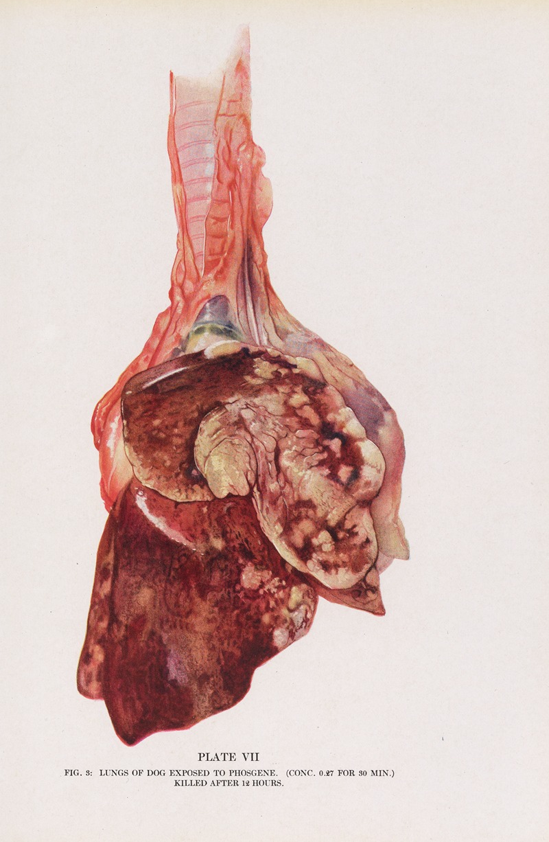
Milton C. Winternitz - Plate VII (Fig. 3): Lungs of dog exposed to phosgene.

Plate VII (Fig. 3): Lungs of dog exposed to phosgene. (1920)

Milton C. Winternitz (American, 1885-1959)
Facebook Twitter Pinterest
Favourite
Collect

Standard, 1174 x 1800px JPG, Size: 1.5 MB

Download

Max Size, 3834 x 5877px JPG, Size: 16.58 MB

Download
License: All public domain files can be freely used for personal and commercial projects.
Why is this image in the public domain?
Courtesy of Science History Institute
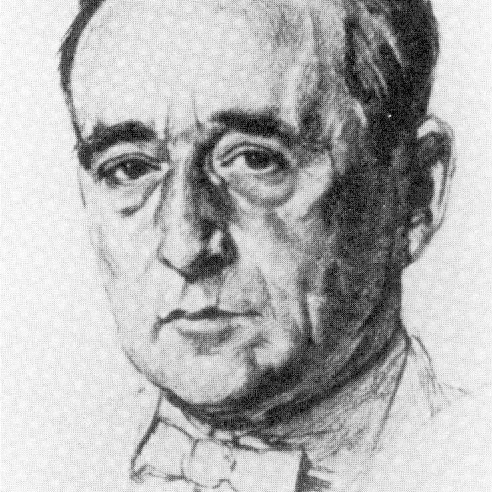
Milton C. Winternitz

Milton Winternitz led Yale Medical School as its Dean from 1920 to 1935. An innovative, even maverick leader, he not only kept the school from going under, but turned it into a first-class research institution. Dedicated to the new scientific medicine established in Germany, he was equally fervent about "social medicine" and the study of humans in their culture and environment. He established the "Yale System" of teaching, with few lectures and fewer exams, and strengthened the full-time faculty system; he also created the graduate-level Yale School of Nursing and the Psychiatry Department, built numerous new buildings, and much more.
More Illustrations in Book: Collected Studies on the Pathology of War Gas Poisoning (View all 30)

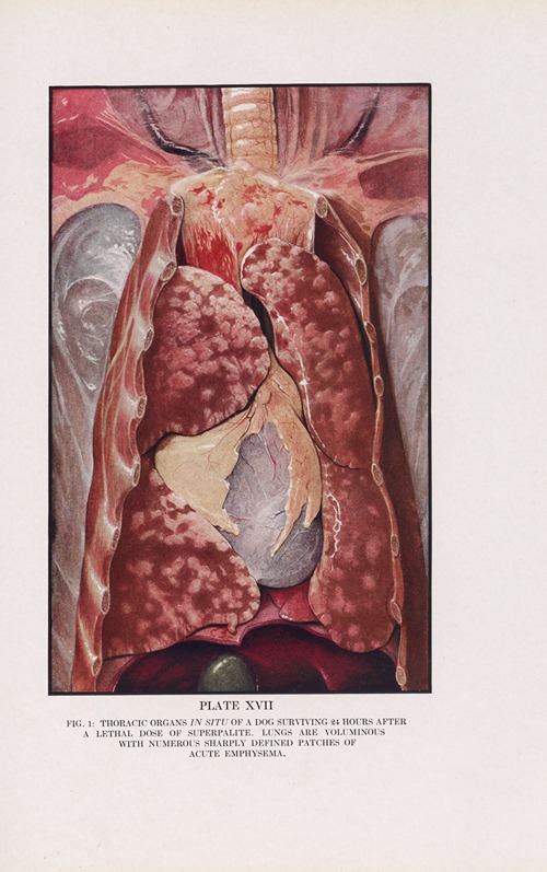
Plate XVII (Fig. 1): Thoracic organs in situ of a dog surviving 24 hours after a lethal dose of superpalite.

Plate XVII (Fig. 1): Thoracic organs in situ of a dog surviving 24 hours after a lethal dose of superpalite. (1920)

Milton C. Winternitz (American, 1885-1959)
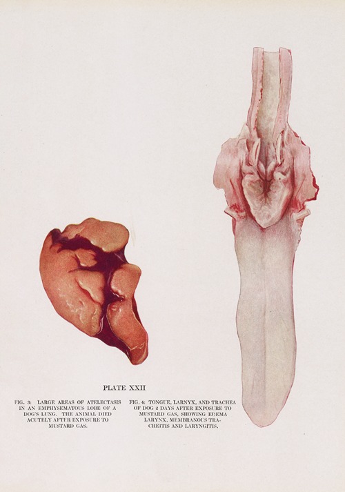
Plate XXII (Fig. 3): Large areas of atelectasis in an emphysematous lobe of a dog’s lung. (Fig. 4) Tongue, larynx, and trachea of dog 2 days after exposure to mustard gas, showing edema of larynx, membranous tracheitis, and laryngitis.

Plate XXII (Fig. 3): Large areas of atelectasis in an emphysematous lobe of a dog’s lung. (Fig. 4) Tongue, larynx, and trachea of dog 2 days after exposure to mustard gas, showing edema of larynx, membranous tracheitis, and laryngitis. (1920)

Milton C. Winternitz (American, 1885-1959)
Plate XXVI (Fig. 22): Eye of dog dying 4 days and 21 hours after exposure to mustard gas. (Fig. 23) Eye of animal dying 7 days after exposure to mustard gas. (Fig. 24) Water color drawing of trachea showing constriction below larynx; 1 month after exposure.

Plate XXVI (Fig. 22): Eye of dog dying 4 days and 21 hours after exposure to mustard gas. (Fig. 23) Eye of animal dying 7 days after exposure to mustard gas. (Fig. 24) Water color drawing of trachea showing constriction below larynx; 1 month after exposure. (1920)

Milton C. Winternitz (American, 1885-1959)
Plate XVIII (Fig. 2): Lungs and trachea of dog dying 2 days after exposure to superpalite.

Plate XVIII (Fig. 2): Lungs and trachea of dog dying 2 days after exposure to superpalite. (1920)

Milton C. Winternitz (American, 1885-1959)
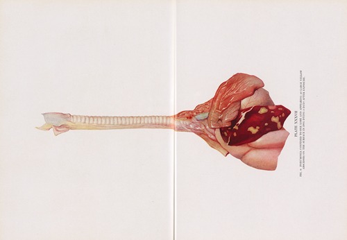
Plate XXXVII (Fig. 6): Pneumonia confined to one lobe and appearing as large yellow abscesses on the surface in dog dying 5 days after exposure.

Plate XXXVII (Fig. 6): Pneumonia confined to one lobe and appearing as large yellow abscesses on the surface in dog dying 5 days after exposure. (1920)

Milton C. Winternitz (American, 1885-1959)
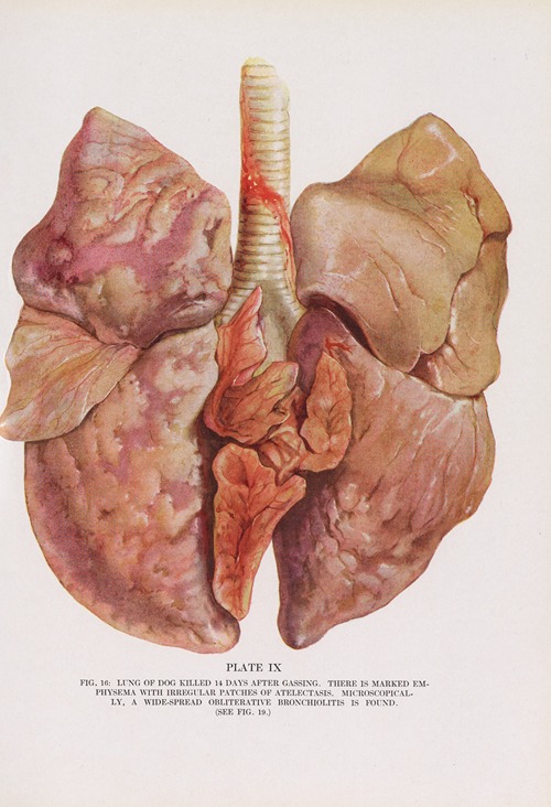
Plate IX (Fig. 16): Lung of dog killed 14 days after gassing.

Plate IX (Fig. 16): Lung of dog killed 14 days after gassing. (1920)

Milton C. Winternitz (American, 1885-1959)
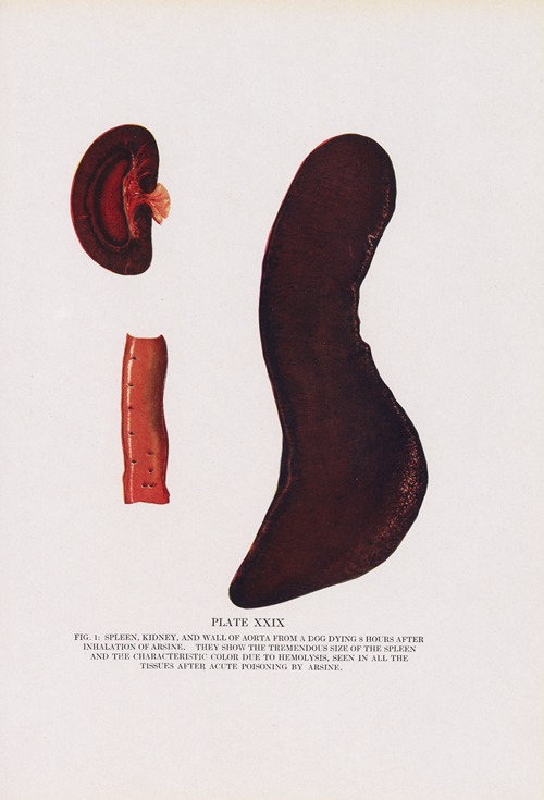
Plate XXIX (Fig. 1): Spleen, kidney, and wall of aorta from a dog dying 8 hours after inhalation of arsine.

Plate XXIX (Fig. 1): Spleen, kidney, and wall of aorta from a dog dying 8 hours after inhalation of arsine. (1920)

Milton C. Winternitz (American, 1885-1959)
Plate XIX (Fig. 8): Lungs of dog surviving 5 days after exposure to superpalite, showing diminution in extent of edema and congestion and nearly complete disappearance of atelectasis and emphysema.

Plate XIX (Fig. 8): Lungs of dog surviving 5 days after exposure to superpalite, showing diminution in extent of edema and congestion and nearly complete disappearance of atelectasis and emphysema. (1920)

Milton C. Winternitz (American, 1885-1959)
Plate V (Fig. 1): Heart and lung of dog dying 24 hours after gassing.

Plate V (Fig. 1): Heart and lung of dog dying 24 hours after gassing. (1920)

Milton C. Winternitz (American, 1885-1959)
Plate XL (Fig. 1): Hemorrhages in lung of dog killed 10 days after phosgene gassing.

Plate XL (Fig. 1): Hemorrhages in lung of dog killed 10 days after phosgene gassing. (1920)

Milton C. Winternitz (American, 1885-1959)
Plate XXV (Fig. 19): Necrotizing membrane of epiglottis and larynx and extensive pneumonia 5 days after a 30-minute exposure to mustard gas.

Plate XXV (Fig. 19): Necrotizing membrane of epiglottis and larynx and extensive pneumonia 5 days after a 30-minute exposure to mustard gas. (1920)

Milton C. Winternitz (American, 1885-1959)
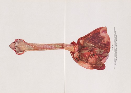
Plate XXXIII (Fig. 1): Trachea and lungs from dog dying 1 day after exposure to ethyldichlorarsine.

Plate XXXIII (Fig. 1): Trachea and lungs from dog dying 1 day after exposure to ethyldichlorarsine. (1920)

Milton C. Winternitz (American, 1885-1959)
Plate VI (Fig. 2): Trachea of same case, opened from behind.

Plate VI (Fig. 2): Trachea of same case, opened from behind. (1920)

Milton C. Winternitz (American, 1885-1959)
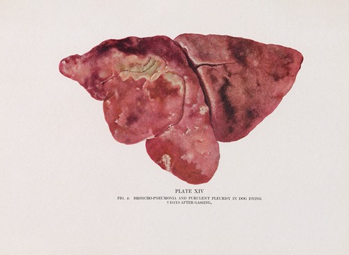
Plate XIV (Fig. 2): Broncho-pneumonia and purulent pleurisy in dog dying 7 days after gassing.

Plate XIV (Fig. 2): Broncho-pneumonia and purulent pleurisy in dog dying 7 days after gassing. (1920)

Milton C. Winternitz (American, 1885-1959)
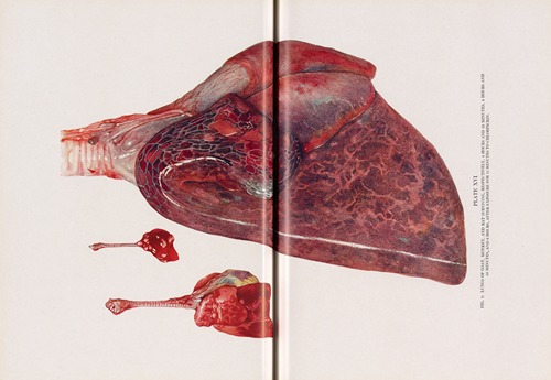
Plate XI (Fig. 2): Lungs of rabbit exposed to phosgene.

Plate XI (Fig. 2): Lungs of rabbit exposed to phosgene. (1920)

Milton C. Winternitz (American, 1885-1959)
View all 30 Artworks

0 Artworks
Follow
Facebook Twitter Pinterest
Favourite
Collect

Standard, JPG, Size:

Download

Max Size, JPG, Size:

Download
License: All public domain files can be freely used for personal and commercial projects.
Why is this image in the public domain?
  • Terms
  • Privacy
  • Contact us
Artvee.com 2024 All Rights Reserved
We use cookies to improve your experience on our website. By browsing this website, you agree to our use of cookies.
More info Accept
  • Sign in
  • Browse
    • Abstract
    • Figurative
    • Landscape
    • Religion
    • Mythology
    • Posters
    • Drawings
    • Illustration
    • Still Life
    • Animals
    • Botanical
    • Asian Art
  • Artists
  • Books
  • Explore
    • Topics
    • Culture
    • Movements
  • Highlights
  • Collections
  • Galleries
  • Artvee Pro